Dental health is an important part of everyone’s life, but unfortunately, it can get expensive. From checkups to treatments, dentistry can involve some pricey procedures — both medical and cosmetic. As with many industries, advancements in dentistry involves turning to newer technologies to reduce time, complexity, and costs. And dental 3D scanners are definitely the way to do that.

Older methods of intraoral inspection are undergoing something of an overhaul, and while the idea of a dental scanner isn’t exactly new, it has seen a few breakthroughs in recent years. This is all in the name of both efficiency, accuracy, and ease, keeping oral health high, and dental bills relatively low.
What Are Dental 3D Scanners?
Otherwise known as mouth scanners, dental 3D scanners scan the shape and form of a patient’s teeth as accurately as possible without being too invasive.
Basically speaking, these scanners take a 3D photograph of the inside of a person’s mouth, converting what they scan into 3D models depicting the shape and contours of the teeth and jaws. These CAD models can then be used to pinpoint problem areas in a patient’s teeth to decide on any necessary procedures.

These procedures can be anything from root canals to complete reconstructive surgeries and braces.
Some scanners are used instead to create 3D scans of existing models like dentures and retainers, converting these scans into .stl files means they can be kept on file, altered, and replicated as necessary to make custom implants or replacements for things like dentures or retainers.
What are the benefits of dental 3D scanners?
The main benefits of dental 3D scanners come down to both efficiency and patient comfort.
While x-rays are commonplace in the world of dentistry, they can only give 2D images to show the outline of a patient’s teeth and highlight problem areas. This combined with a manual inspection is how most dentistry is done.

With intraoral 3D scanners, scans of every angle of the mouth are taken and modeled in only a minute, with detailing close enough to reality to be used in making diagnoses, deciding on procedures, and measuring dimensions for things like implants, dentures, and retainers.
Faster Analysis
Dental professionals and lab technicians are not always the same thing. There are always far more people working behind the scenes of medical practices than we often realize, and communication between the departments that diagnose and those that prepare treatments is imperative to a patient’s health.
Digital dental scans can be shared between these departments instantaneously. Instead of running x-rays and notes around a practice, a scan can be converted into a file that is then sent to the lab along with any relevant notes.
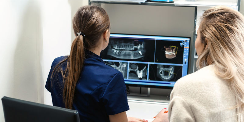
From there, the lab can begin immediately on analyzing and using the scan to begin creating prosthetics. Lab technicians and active practitioners need to have as fluid and as streamlined a communication system as possible, and digital dental scans make such communication easy, efficient, and very fast.
Patient Comfort
Older methods for measuring out the contours of a patient’s teeth mainly involved rather invasive methods, including molds being made with a silicone-like substance. This substance is layered over the teeth to make an impression, similar to pressing Silly Putty on something to form its outline.
This is fairly uncomfortable and invasive, but necessary for making retainers and dental implants. With a handheld intraoral 3D scanner, this discomfort is replaced with a relatively fast internal inspection of the mouth, scanning the teeth and making an accurate 3D model representing the shape of the patient’s teeth.

This process is not only more comfortable for the patient, but is also far faster and more efficient. Without the need to physically make a mold of the teeth, a process that needs to be repeated as the shape of someone’s teeth changes via age or lifestyle, a CAD file can be altered to suit the new shape before being sent to print.
As an added bonus, intraoral and digital scanners don’t use the kind of radiation that x-rays do, making them safer and less likely to cause any damage in the long run. This is especially true for patients who need frequent x-rays, which can not only increase the risk of cancer, but can also have quite nasty short-term side effects.
While the risks of such side-effects (vomiting, hair loss, numbness) are admittedly low, the reduction of any risk at all by replacing x-ray imaging with 3D scanners makes them safer.
Speed
With dental 3D scanners, scanning a patient’s mouth repeatedly over the course of treatment takes very little time and relatively few materials, with most scans being completed from scan to model in as little as 20 minutes.
The speed of the scans, when combined with dental 3D printing, means that custom aligners, retainers, and dentures can be made on the same day. This reduces wait times, removes the need for return visits, and comes at a much lower cost to both dentists and patients as they can be made quickly in-house with no transporting.

This speed not only saves time and money for dentists and patients, but also ensures that emergency procedures or time-sensitive treatments can be carried out quickly. This promotes earlier and better healing as treatments can be provided before a condition worsens.
Efficiency
For more complicated surgeries, larger 3D scanners can be used alongside x-rays to manifest a full representation of a patient’s entire jawline. This aids in procedures like facial reconstruction and resetting of broken bones in far less time than was possible before. A standard x-ray can take up to 7 minutes, while a dental scan can take as little as 20 seconds.
These larger scans are carried out with 3D scanners similar to an x-ray that rotate around your head and constantly capture pictures, much like a video camera. The reduced need for physical inspections is much better for patients, more accurate for practitioners, and far more reliable for the lab techs.
Education
As well as being great for active dental practices, digital scanners make for excellent teaching tools. By creating accurate models of a patient’s teeth, students can learn how to best identify problems and go about any necessary procedures in practical, even interactive, ways.
This is much like how visual representations of camera angles aid media students, or how attending autopsies helps medical students grow their in-depth knowledge of anatomy; analyzing 3D scans of real teeth are a great way for dental students to get further insight into what to expect from their future jobs.
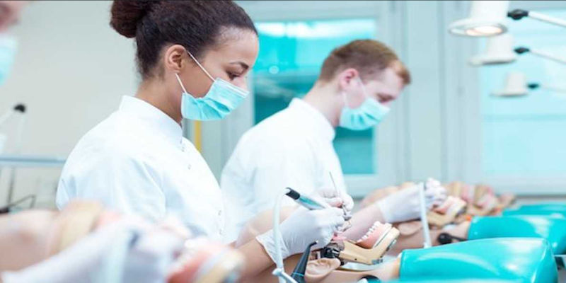
This kind of education isn’t only useful for pre-medical learning, but also for actively training new dentists in internships and placements. As technology evolves for professional use, early issues often involve workers being undertrained.
Using scans taken with lab scanners, those still in training can also simulate how they would alter an implant or other prosthetic in a controlled environment that wastes neither material nor money in the event of failure.
What are the Best Dental 3D Scanners Available?
Dental scanners come in many forms, and like most examples of medical technology, the different kinds each have their own specific uses.
Here, we’re going to look at the most common types of intraoral scanners in use today.
Intraoral 3D Scanners
Intraoral Scanners are handheld devices that look like large electric toothbrushes. They’re typically light, fast, and accurate, though can’t provide as detailed a scan as some of the larger scanners.

The Planmeca, for example, is a handheld 3D dental scanner model that’s held in high esteem for its speed and accuracy, especially for a device with such a small and lightweight design. The Planmeca Emerald S is an upgraded version, faster and more accurate than its predecessor. The Emerald S is also designed to work with multiple file types for easy sharing across different CAD and CAM software.
While primarily designed for adult scans, smaller tips are often available for younger patients or patients with smaller mouths than average. This is useful for scanning anterior teeth with greater accuracy than a larger tip would allow.
Cone-Beam Computed Tomography
Cone-Beam computed tomography acts more like a 360° x-ray than a standard blue light scanner. Small cones of x-ray radiation are applied in a sort of helmet, and a full 3D scan of a patient’s teeth is taken without the patient ever needing to open their mouth.
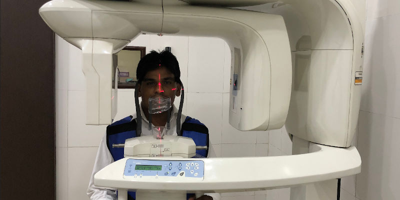
This non-invasive scanning method may use x-rays, but is still far safer and exposes patients to much less radiation than a traditional x-ray.
The key advantage is the automatic motion correction these devices provide, similar to how most modern camera phones will adjust for any small movements to get as clear and accurate an image as possible.
3D Dental Lab Scanners
3D dental lab scanners are similar to the kind of scanners we can get on our smartphones, and are used mainly for restorative surgeries and prosthetics like dentures and implants.
The scans made with a lab scanner can be kept on file for future prosthetics as necessary, or used to 3D print corrections to any existing prosthetics or orthodontics.

Most lab scanners will export as STL files to be used in 3D productions, though they don’t act as internal scanners and are mainly used for gypsum models and impressions. The scans can then be used to create replicas for spare dentures or retainers, or used to make alterations should a patient’s condition change.
Lab scanners are by far the fastest scanners on the market and can perform a full arch scan in as little as 8 seconds. This is very impressive when compared to smaller, handheld devices like intraoral scanners, which can take around a minute for a full mouth scan.
Other articles you may be interested in:

